Magnetic Resonance Imaging
Magnetic Resonance Imaging
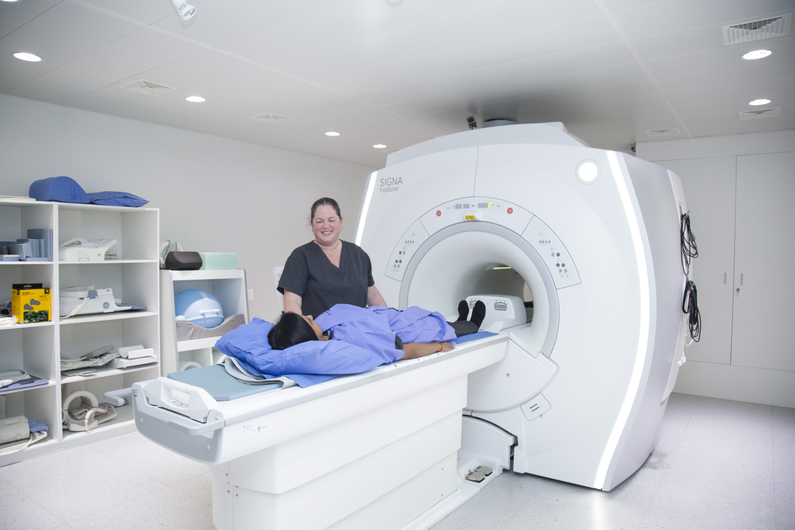
MRI or Magnetic Resonance Imaging, uses a powerful magnet and radio waves to produce superbly detailed views of the human body, particularly soft tissues, such as the brain, spinal cord and muscles. Unlike other imaging tests, MRI does not use radiation to acquire the images.


- Please remember to bring your medical aid card together with the signed request form from your referring doctor.
- Medical aid authorisation will be obtained by our department.
- A motivation letter may be required from your doctor, which can delay your authorisation.
- Co-payments may apply to certain medical aids, this will be discussed at the time of booking. Payment is due at the time of the scan and is your percentage contribution of the rate that medical aids pay the radiologist. For example medical aid payment =R6000 – your co-payment R2700 Medical aid pays towards your scan R4300.
- An appointment time will be given to you; however, if there are delays with the authorisation, we will let you know.
- Please arrive 20 to 25 minutes before the appointment time to complete paperwork.
- Previous imaging, not done with Lake, Smit & Partners, must be brought with you to the scan.
- You will be asked to read and complete an MRI Consent and Safety Questionnaire. This will be checked by the Radiographer before your scan.
- Due to the strong magnet within the MRI, we will ask that you remove all jewellery, cell phones, wallets and anything else metallic. A lockable cupboard is available in the change cubicle.
- A cotton gown will be provided for you to wear during the examination.
- The examination takes approximately 20 minutes to 1 hour, depending on the examination.
- Please empty your bladder before the scan.
- Due to the enclosed space within the MRI Scanner light sedation may be recommended. If you require this, please arrange for a responsible driver to fetch you and refrain from driving and using any machinery for the rest of the day.
Please inform your Radiographer before the scan if any of the following apply to you:
- A non-MRI compatible Aneurysm clip;
- A non-MRI compatible Pacemaker;
- Certain electronic/metallic implants;
- Certain cochlear implants;
- Bullets or shrapnel close to vital organs;
- Certain eye implants or foreign bodies;
- Female patients, should you be, or suspect you may be pregnant.
Please bring along any information regarding your implant. As a general rule, MRI scans are usually not conducted in the first trimester of pregnancy unless it is deemed by the referring doctor or radiologist that is an absolute medical necessity to do so and that the benefits of the test outweigh the risks. This of course assumes that there is no other test available that can provide similar information.
What to expect during the MRI Scan
- You will be asked to lie on a movable scanning table.
- During the MRI examination, the scanner emits a repetitive noise and you will be given earplugs to ensure your comfort.
- Due to the loud noises of the scanner, you will be offered earplugs or headphones.
- A device, known as a coil, will be placed over the area of examination. You will then be moved to the middle of the scanner.
- The Radiographer will be able to communicate with you at all times. A handheld alarm buzzer will be given to you at the start of the scan to ensure your safety.
- Some examinations may require an IV injection of contrast media. This will be given halfway through the scan.
MRI Contrast (Gadolinium)
Some patients undergoing an MRI scan may require an injection of an intravenous dye (contrast) known as Gadolinium, which enhances pathology and tissue detail on your scan. The contrast is delivered into your body through an intravenous cannula, which is placed into a vein in your arm by a nurse or radiographer who are both experienced in performing this procedure.
The decision to administer IV contrast is not taken lightly and is carefully made by the radiologist and is based on your signs, symptoms, past medical history as well as the suspected diagnosis.
Most injections of IV contrast occur uneventfully. The most common side effect is a minor contrast reaction, which occurs in less than 0.05% of cases. Symptoms include headache, sneezing, nausea, vomiting, hives and swelling and usually settle rapidly. Occasionally medications may be required to help alleviate the symptoms if they persists for some time and typically involve using an anti-emetic and promethazine (phenergan) for urticarial and swelling. Phenergan unfortunately results in drowsiness, so is usually administered only if the swelling or hives is particularly severe. In this instance, you would need a responsible person to drive you home.
- Less commonly, a severe (anaphylactoid) contrast reaction occurs in approximately 0.03–0.1% of cases. This includes a rapid or slow
- heart rate, low blood pressure, an asthma attack (bronchospasm) and rarely complete circulatory collapse/shock. Such reactions require urgent medical treatment and immediate transfer to an appropriate facility, such as an emergency department or intensive care unit. All our departments have a fully equipped emergency trolley to ensure appropriate medical treatment.
- Patients with kidney (renal) impairment or failure should not undergo an injection of gadolinium unless this has been cleared by a specialist in this field (renal physician) in order to avoid a very rare but potentially life threatening condition known as NSF (Nephrogenic Systemic Fibrosis).
- Patients who have had a contrast reaction to the dye used in CT, IVP and angiographic examinations are at a 3.7 times increased risk of an adverse reaction.
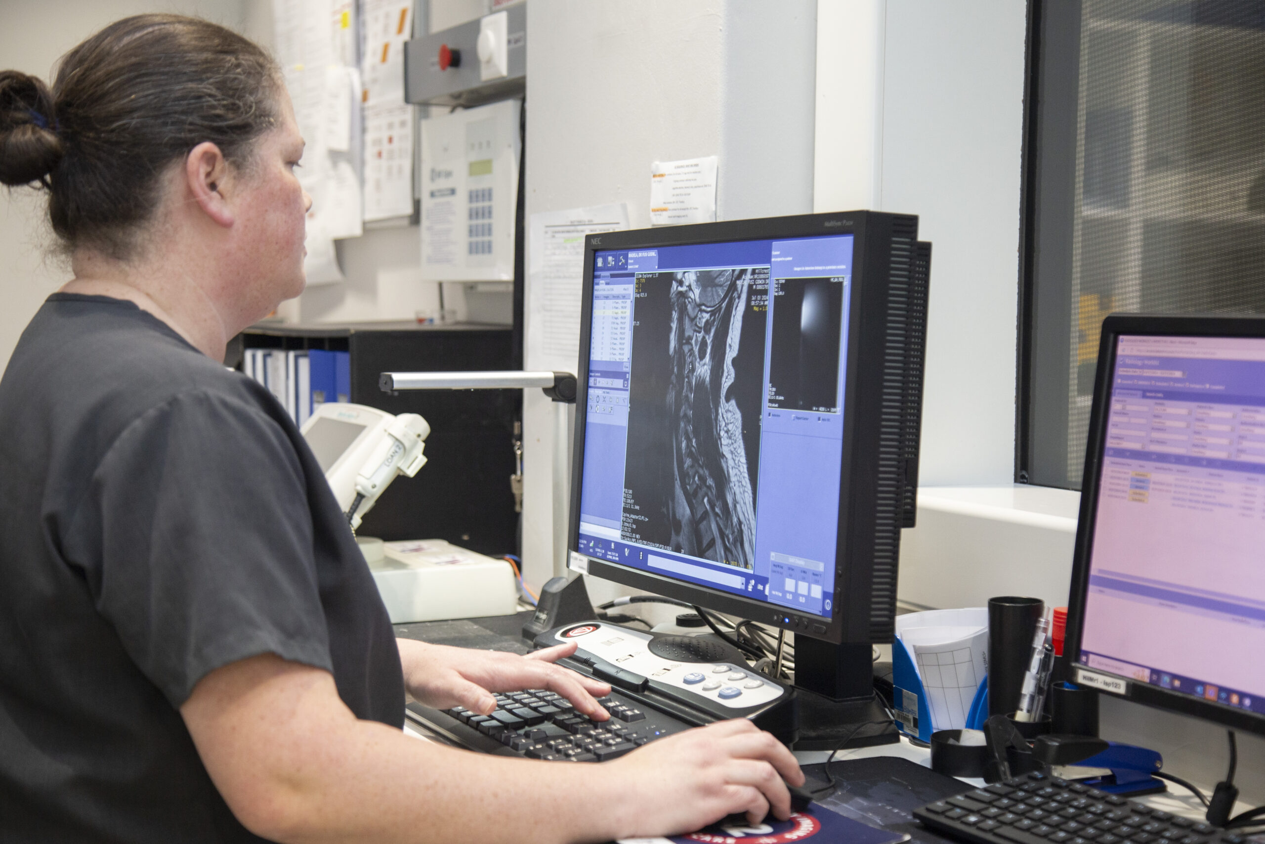
These examinations require interpretation from a Specialist Radiologist that may take any time between a few hours up to 24 hours for complex studies. Once this is completed, your report will be sent to your referring doctor.
St Augustine’s Hospital
107 JB Marks Road, Durban
Tel: 031 521 0374
Entabeni Hospital
148 Mazisi Kunene Road, Durban
Tel: 031 521 0373
Westville Hospital
7 Harry Gwala Road, Westville, 3630
Tel: 031 521 0372
Gateway Private Hospital
36-38 Aurora Drive, Umhlanga Rocks
Tel: 031 521 0375
Hillcrest Imaging Centre
D1 Meyrickton Park
Tel: 031 521 0380

