3D MAMMOGRAPHY

3D MAMMOGRAPHY

A mammogram is a low dose x-ray image done of your breasts using compression to allow for equal penetration of the x-ray beam through the breast tissue, creating detailed images of your breasts.

It is considered the gold standard for early detection of breast cancer, capable of identifying abnormalities such as masses, distortions or microcalcifications that may not be detectable through physical examination alone. Mammography is highly effective, often detecting breast cancers long before they can be found by clinical examination, ultrasound, or other methods.

Breast tomosynthesis, also known as 3D mammography, is an advanced imaging technique offered at most mammography centres these days. It captures multiple detailed images from various angles, enabling radiologists to examine breast tissue layer by layer. This improves the detection of small subtle cancers, resulting in more precise diagnoses and earlier identification of breast cancer.

  • Lower radiation does
  • Superior image quality
  • Εαrly detection
  • Improved cancer detection rate
  • Softer, more gently mammogram

Just for Women Digital Mammography Centre:

  • Full-time Specialist Radiologists and Mammographers;
  • Women’s Health & Preventative Care all under one roof (this includes Mammography, Ultrasound & Bone Densitometry);
  • A stand-alone centre, that’s not in a hospital environment;
  • Dedicated parking;
  • A tea lounge, to enjoy a cup of tea or coffee after your mammogram in a relaxed environment.
  • Please remember to bring your medical aid card together with the signed request form from your referring doctor.
  • You must get authorisation from your Medical Aid before your appointment.
  • An appointment time will be given to you.
  • You can book your mammogram by emailing central.bookings@lakesmit.co.za

Before your Mammogram

  • Please arrive 10 to 15 minutes before your appointment time to complete the paperwork.
  • Previous imaging, not done with Lake, Smit & Partners, must be brought with you to the scan.
  • You will be asked to complete a questionnaire before your mammogram is done.
  • A cotton gown will be provided for you to wear during the examination.
  • Do not wear any talcum powder or deodorant on the day of your examination.
  • The examination takes approximately 15 minutes.

Are mammograms safe?

  • The benefits of mammography outweigh any possible harm from the radiation exposure. Modern machines use low radiation doses to get breast x-rays that are high in image quality. On average the total dose for a typical mammogram with 2 views of each breast is about 0.4 mSv. (A mSv is a measure of radiation dose.)
  • To put dose into perspective, people in the US are normally exposed to an average of about 3 mSv of radiation each year just from their natural surroundings. (This is called background radiation.) The dose of radiation used for a screening mammogram of both breasts is about the same amount of radiation a woman would get from her natural surroundings over about 7 weeks.
  • If there’s any chance you might be pregnant, let your health care provider and x-ray technologist know. Although the risk to the fetus is likely very small, screening mammograms aren’t routinely done in pregnant women.

Why not just an ultrasound?

  • Ultrasound is limited as a screening method as it may not detected small areas of abnormality but is excellent as a examination directed to an abnormality seen on mammogram.
  • However in patients younger than 40 ultrasound is the method of choice to evaluate an abnormal area in the breast.

During your Mammogram

  • 4 images will be taken of your breasts.
  • You will be standing while the X-rays are taken.
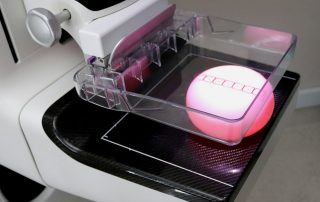
  • Your breast will be placed on a plate and compressed by a second perspex paddle (good positioning and the compression technique is vital to ensure we get a high quality image).
  • The X-ray unit will move above your head while the images are taken.

After your Mammogram

Sometimes an ultrasound is needed to visualise any abnormalities in the breast tissue. If this is the case, it will be arranged at the time of the Mammogram. If there is any reason for concern, our Radiologist and Mammographers will discuss this with you and any further investigations that are necessary will be explained to you and then booked.

Just For Women
T Medical Centre 2nd Floor
22 Hopelands Road Overport
Tel: 031 521 0379

Just for Women Digital Mammography Centre:

  • Full-time Specialist Radiologists and Mammographers;
  • Women’s Health & Preventative Care all under one roof (this includes Mammography, Ultrasound & Bone Densitometry);
  • A stand-alone centre, that’s not in a hospital environment;
  • Dedicated parking;
  • A tea lounge, to enjoy a cup of tea or coffee after your mammogram in a relaxed environment.

The benefits of 3D Mammography:

  • Lower radiation dose
  • Superior image quality
  • Early detection
  • Improved cancer detection rate
  • Softer, more gentle mammogram

CONTACT US TODAY

You can contact us by using our contact form below. We look forward to hearing from you.

REMEMBER! Early detection is the best detection, every time! Breast cancer can be successfully treated when it’s detected early.